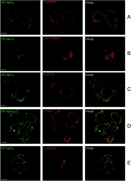
FIG. 6.
Confocal analysis of the distribution of TAT-NpFlu chimeric protein. TAT-NpFlu fusion protein is visualized in green (left panels) and cellular compartments are depicted in red (middle panels); the overlays are shown on the right. (A) Transferrin, the marker for early endosomes. (B) Lysotracker, marker for late endosomes and lysosomes. (C) Anti-CD-63 for lysosomes and multivesicular bodies. (D) Anti-caveolin-1 (Cav-1) for caveolar compartments. (E) Anti-Giantin for cis and medium Golgi. TAT-NpFlu is marked with FITC-conjugated secondary antibody. Bars, 10 μm.

