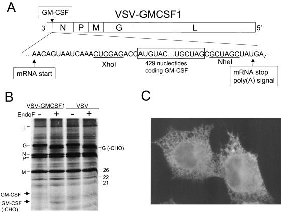
FIG. 1.
Recombinant VSV genome and protein expression. Panel A is a diagram showing the gene order of the VSV-GMCSF1 recombinant. The gene order is shown in the 3′-5′ direction of transcription on the negative-strand RNA genome. The GM-CSF gene insertion site and flanking nucleotides, including the transcription and translation start and stop sites, are indicated. Restriction enzyme sites used for cloning the GM-CSF gene at the DNA stage are also indicated. All sequences are shown in the positive (antigenome) sense for clarity. Panel B shows an SDS-PAGE (15% acrylamide) gel containing lysates of BHK cells infected with the indicated viruses and labeled with [35S]methionine. The gel image was collected by autoradiography on X-ray film. Panel C shows an immunofluorescence image of BHK cells infected with VSV-GMCSF1. Cells were infected for 4 h, fixed, permeabilized, and stained with a PE-labeled monoclonal antibody recognizing mGM-CSF. Cells were photographed using a Nikon fluorescence microscope with a 60× Planapochromat objective and a SPOT digital camera.

