FIG. 9.
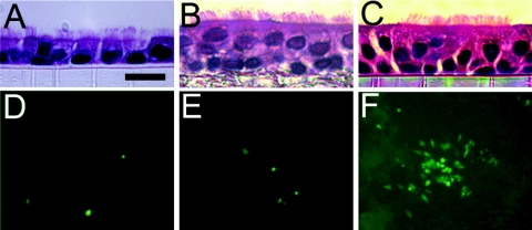
SARS-CoV GFP infection of mouse, hamster, and rhesus monkey airway epithelial cell cultures. Ciliated airway epithelial cell cultures were derived from mice (A and D), hamsters (B and E), and rhesus monkeys (C and F) and inoculated via the apical surface with SARS-CoV GFP (106 PFU). Hematoxylin and eosin-stained histological sections are shown for each species (A, B, and C), and GFP fluorescent images were recorded 48 h postinfection (D, E, and F). Original magnifications were ×40 (A, B, and C) and ×10 (D, E, and F). Bar, 5 μm (A, B, and C).

