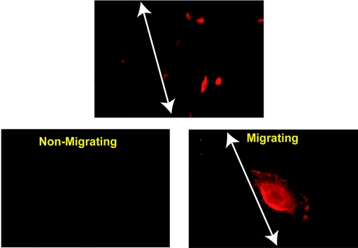
FIG. 6.
ITGα6 expression is up-regulated in migrating primary epithelial cells. A scratch wound assay with adLMP2A-infected primary epithelial cells was performed on culture slides as described for Fig. 5. The cells were then fixed and incubated with an ITGα6-specific antibody (GoH3), and ITGα6 expression was visualized by indirect immunofluorescence with an Alexa-594 conjugated goat anti-mouse secondary antibody (red). In the top (low magnification) and bottom right (high magnification) panels one edge of the scratch is delineated with a white arrow. The unaffected monolayer of epithelial cells is to the left of the line. The lower left panel shows a field of nonmigrating cells from an area of the culture away from the scratch to demonstrate the staining obtained with confluent cells. Note that ITGα6 staining is weak with the unaffected monolayer but that strongly positively stained cells are only observed migrating into the scratch to the right of the white arrows. The upper image was taken at ×40, and the lower two images were taken at ×100 magnification.

