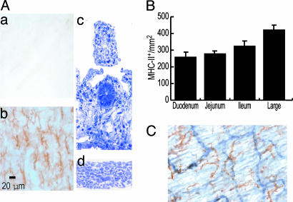
Fig. 1.
A dendritic, strongly MHC-II+ population in the intestinal layer toward the serosa. (A) The intestine was incubated in 0.5 M EDTA for 30 min at 37°C, two layers were separated by traction by using fine forceps, and the layers were fixed in cold acetone. (a and b) Upon labeling, MHC-II+ cells with clear dendritic morphology and a regular pattern of distribution are observed in only one of the two intestinal layers obtained (toward the serosa). To study the precise histological location of this DC population, transverse semithin sections of each layer were obtained. The layer (c) toward the lumen comprises the lamina propria and submucosa, whereas the layer (d) toward the serosa corresponds to the external muscular layer. (B) The frequency of this MHC-II+ population was also analyzed in each of the different anatomical regions of the intestine. (C) The possible association of the MHC-II+ (brown) DC to vessels was assessed by double labeling for CD31 endothelial molecule (blue). The MHC-II+ DCs do not appear associated with CD31+ (blue) vessels (blood or lymphatics).

