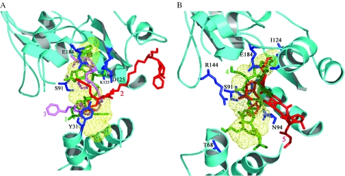
Figure 2.
(A) Interactions of compounds 1, 2 and 3 (those with aminoalkyl spacers) in the NAD+ binding site of the enzyme from M.tuberculosis. The compounds are shown in green, red and pink respectively while the NAD+ binding region is depicted as a light green wire mesh. Key interacting residues are shown as blue sticks and labeled for clarity. (B) Interactions of compounds 4 and 5 (those with phenylene carbamoyl spacers) in the NAD+ binding site of the M.tuberculosis enzyme. The compounds are depicted in green and red, respectively. The color scheme is similar to (A).

