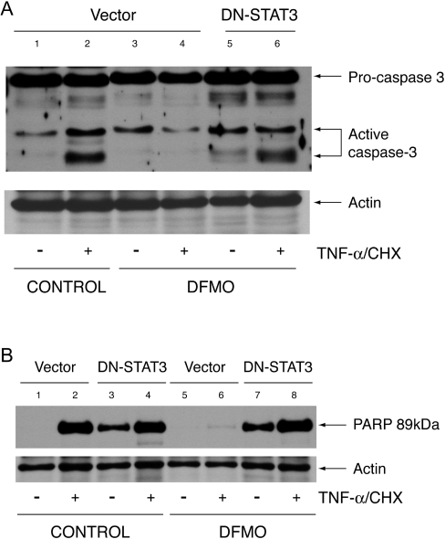
Figure 9. Caspase-3 activation and PARP cleavage.
Cells transfected with vector and DN-STAT3 were grown as described in the Experimental section and treated with TNF-α/CHX or left untreated. Protein (40 μg) from each sample was separated by SDS-gel electrophoresis, transferred to membrane and probed with caspase-3 antibody (A) and a cleaved PARP (Asp-214) rat-specific antibody (B). Membranes were stripped and probed with β-actin antibody as an internal control for equal loading. Representative blots from three experiments are shown.

