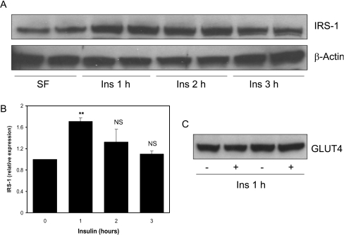
Figure 3. Induction of IRS1 protein by insulin in L6 myotubes.
(A) Rat L6 myotubes were serum-starved overnight and treated with 100 nM insulin for 1, 2 or 3 h. Western blot analysis was performed on 50 μg of cell lysates to assess IRS-1 and β-actin protein levels. (B) Expression was quantified by densitometric scan of the blots. Values are the ratio of IRS1 to β-actin and are presented relative to serum-starved cells (average±S.E.M. of two experiments performed in duplicate). **P<0.01 (0 compared with 1 h); NS, not significant. (C) GLUT4 extraction following L6 cell lysis was assessed by Western blot analysis.

