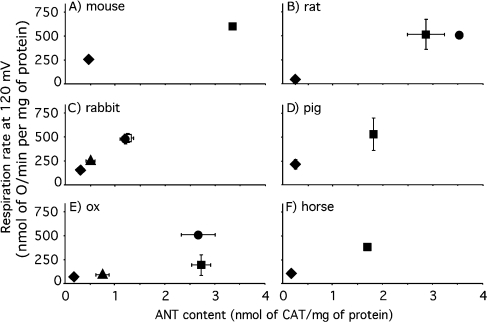
Figure 10. Correlation between proton conductance and ANT content in mammalian mitochondria.
Mitochondria were isolated from liver (◆), skeletal muscle (■), heart (●) and kidney (▲) of the species indicated. The dependence of proton-leak rate (measured as the respiration rate driving proton leak in the presence of oligomycin) on membrane potential was determined as in Figure 3(A) and the interpolated rates at the highest common potential (120 mV) were calculated using mean data. Error bars represent the weighted mean of the respiration rate S.E.M. (or range when n=2) values of the flanking experimental points. ANT content was determined by CAT titre as in Figure 1(B); values are means±S.E.M. (or range when n=2). The numbers of independent preparations were one (mouse and horse tissues, and rat heart), two (pig tissues, rat liver and ox heart), three (ox liver, kidney and skeletal muscle) or four (rabbit tissues and rat skeletal muscle).

