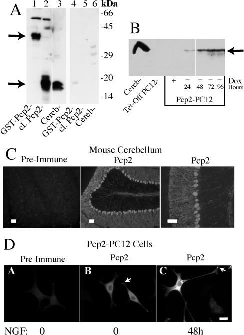
Figure 1. Establishment of Tet-off inducible Pcp2 expression in PC12 cells.
(A) Western blot demonstrating sensitivity and specificity of Pcp2 antibody. Lanes 1–3 show detection of Pcp2 at 1:5000 (0.4 μg/ml) dilution and lanes 4–6 are duplicate samples analysed by Western blotting after preincubation of the antibody with a 3-fold molar excess of GST–Pcp2 protein. Lanes 1 and 4, GST–Pcp2 (50 ng); lanes 2 and 5, cleaved (cl.) Pcp2 [by thrombin cleavage of GST–Pcp2 protein (100 ng)]; and lanes 3 and 6, mouse cerebellar lysate (75 μg). (B) Western blot of Pcp2 expression in Tet-off transfected PC12 cell line. Dox at 50 ng/ml (+) for 96 h was compared with parallel cultures grown in the absence of Dox for 24, 48, 72 and 96 h. Cerebellar lysate (100 μg) is shown in the first lane. 50 μg of PC12 lysate was used in each lane and Pcp2 antibody at 1:1000 dilution. (C) Pcp2 staining in mouse cerebellum. Frozen sections were fixed and stained with Pcp2 antibody at 1:1000 dilution or preimmune serum as described in the Experimental section. Scale bar=100 μM. (D) Immunolocalization of Pcp2 in PC12 cells cultured −Dox for 48 h. PC12 cells were grown on poly-lysine coated glass coverslips and stained with Pcp2 antibody or preimmune serum at 1:500 dilution as described in the Experimental section. Pcp2 localization under basal conditions (no NGF) was compared with Pcp2 expressing PC12 cells differentiated for 48 h with NGF (B, C). Images were obtained using a Nikon Labophot-2 microscope and Spot Digital camera and software (http://www.diaginc.com/SpotSoftware; version 3.5.7) and Figures assembled using Adobe Photoshop and Illustrator. Scale bar=10 μM.

