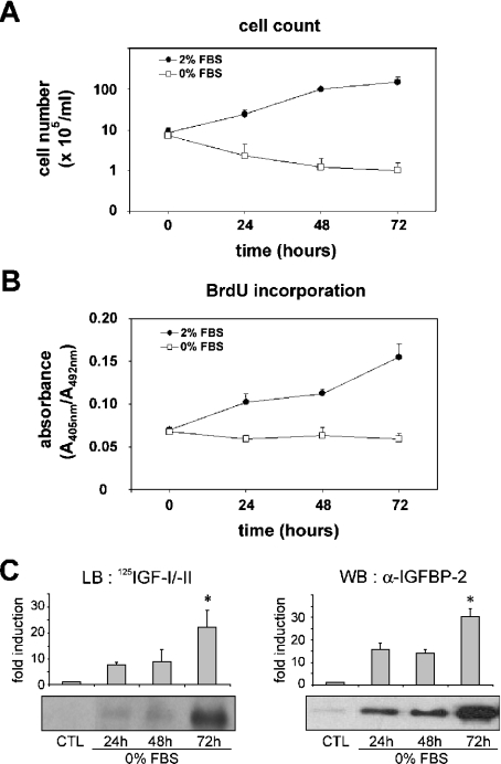
Figure 1. Effect of serum starvation on proliferation of MLE-12 cells and IGFBP-2 secretion.
Sub-confluent MLE-12 cells were cultured in HITES medium for 24, 48 or 72 h in the presence (●) or absence (□) of serum. (A) Cell counts were determined per dish using Trypan Blue exclusion assay. (B) DNA synthesis was determined as arbitrary units of absorbance using BrdU incorporation assay. (C) Ligand blot and Western blot analysis of secreted IGFBP-2. Samples from conditioned media corresponding to 3×10⁵ cells were desalted, lyophilized, resuspended and analysed by SDS/PAGE. IGFBP-2 capacity for binding IGF was evaluated by Western ligand blotting (LB) by binding with 125I-IGF-I and 125I-IGF-II (left panel). There was a 32 kDa band corresponding to IGFBP-2, which was confirmed by Western blotting (WB) with anti-IGFBP-2 antibody (right panel). Histograms show a quantitative representation obtained from laser densitometric analysis of three independent experiments. Densitometry results were expressed as fold induction. *P<0.05 compared with control (CTL).

