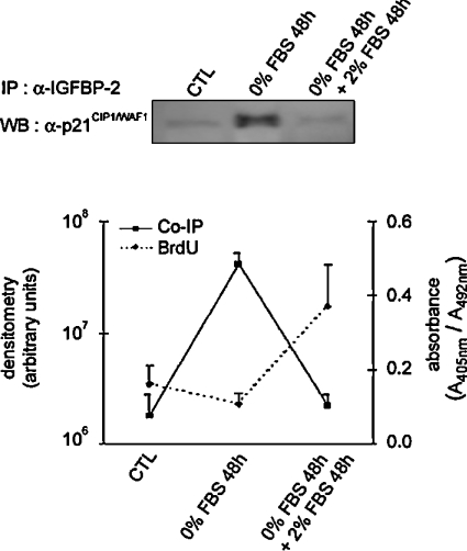
Figure 5. Effect of serum addition to serum-starved MLE-12 cells on proliferation and on the interaction between IGFBP-2 and p21CIP1/WAF1.
Sub-confluent MLE-12 cells were seeded and cultured in HITES medium for 24 h in the presence of serum (CTL), then serum-starved for 48 h (0% FBS 48 h), before the addition of serum for 48 h (0% FBS 48 h+2% FBS 48 h). Total protein extracts from cultured MLE-12 cells were immunoprecipitated (IP) with anti-IGFBP-2 antibody and p21CIP1/WAF1 was detected with anti-p21CIP1/WAF1 antibody in the precipitated complex on a Western blot (WB). A representative blot is shown above the graph. BrdU incorporation was assayed at the three time points (as outlined in the Experimental section). Densitometry from coimmunoprecipitation experiments and absorbance from BrdU incorporation, each representative of three independent experiments, were plotted as a continuous line and a dashed line respectively.

