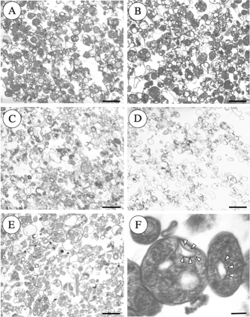
Figure 6. Representative electron micrographs of the subcellular fractions of epimastigotes.
Note that reservosomes are enriched in B1 (A) and B2 (B) fractions. B3 (C) and B4 (D) contain other vacuoles that resemble mitochondrial fragments and microsomes, whereas the pellet (E) fraction also contains flagella and acidocalcisomes. (F) Shows reservosomes of fraction B1 at higher magnification showing the presence of internal membrane-bound vesicles (arrowheads). Scale bars, 0.5 μm (A–E) and 0.1 μm (F).

