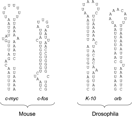
Figure 8. Comparison of secondary structure of the localization elements in K-10, orb, c-myc and c-fos mRNAs.
Structures shown are those proposed for the Drosophila K-10 and orb mRNAs [24] and those for mouse c-myc and c-fos determined in the present study by a combination of chemical cleavage and computer prediction using Mfold.

