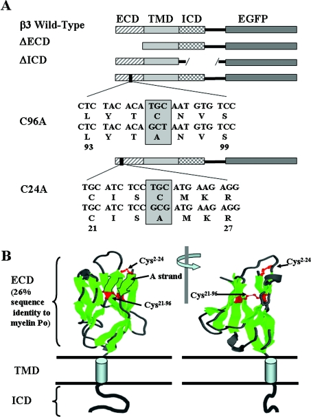
Figure 1. Summary of the mutant constructs.
(A) Diagramatic representation of the EGFP-tagged β3 constructs showing the relative positions of the ECD, TMD and ICD. The ΔECD mutant had amino acids 1–135 removed. The ΔICD mutant had amino acids 158–191 removed. Point mutations C24A and C96A in the ECD are indicated. (B) Modelling of the V-type Ig fold of the ECD. The representation is based on the known structure of myelin P0, a protein showing approx. 26% amino acid sequence identity with the ECD of β3 [9] and for which an accurate structure is known [25]. The model shows the proposed location and disulphide bonds for Cys2–Cys24 and Cys21–Cys96.

