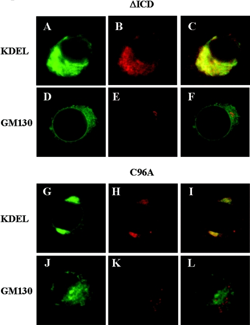
Figure 3. Intracellular localization of ΔICD and C96A mutants.
(A–F) The EGFP-tagged β3 ΔICD and (G–L) the EGFP-tagged C96A mutants were transiently transfected into PC12 cells. (A, D, G, J) EGFP staining. (B, H) Cells co-stained with Cy3-labelled antibodies (red) raised against KDEL, a marker for soluble proteins of the ER. (E, K) Cells co-stained with Cy3-labelled antibodies (red) raised against GM130, a marker for the cis-Golgi network. (C, F, I, L) Merged images.

