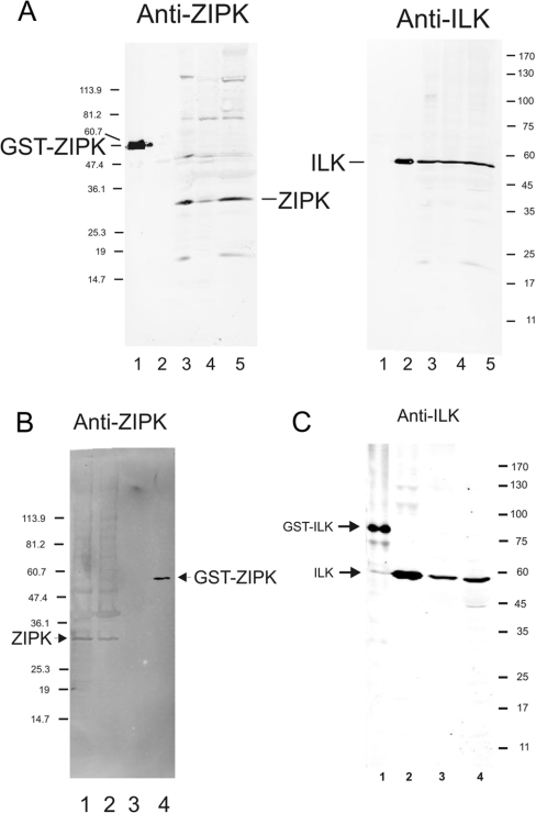
Figure 2. ZIPK and ILK are retained in Triton-skinned rat caudal artery.
(A) Western blots probed with anti-ZIPK (left panel) or anti-ILK (right panel): GST–ZIPK (lane 1), chicken gizzard ILK (lane 2), rat bladder (lane 3), rat aorta (lane 4) and rat caudal artery (lane 5). (B) Western blot probed with anti-ZIPK: intact rat caudal artery (one strip; lane 1), Triton-skinned rat caudal artery (one strip; lane 2), chicken gizzard ILK (lane 3) and GST–ZIPK (lane 4). (C) Western blot probed with anti-ILK: GST–ILK (lane 1), chicken gizzard ILK (lane 2), Triton-skinned rat caudal artery (one strip; lane 3) and intact rat caudal artery (one strip; lane 4). Numbers at the right or left of each panel indicate the sizes of molecular-mass markers (in kDa).

