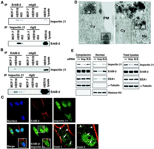
FIG. 1.
ErbB-2 interacts with importin β1. (A) Lysates of the cytoplasmic fraction from MCF-7, MCF-7/HER18, and MDA-MB-453 cells were immunoprecipitated (IP) with antibodies against ErbB-2, importin β1, control mouse IgG (mIgG), and rIgG. The presence of importin β1 and ErbB-2 in the immunocomplexes was examined by immunoblotting analysis. Total cell lysate from the MCF-7/HER18 cells was used as the positive control. (B) Nuclear lysates from the same cell lines were tested for the association between ErbB-2 and importin β1 as described for panel A. Total cell lysate from the MCF-7/HER18 cells was used as the positive control. (C) ErbB-2 and importin β1 colocalized in the cytoplasm (insets 1 and 2, arrowheads) and the nucleus (insets 1 and 2, arrows) of MCF-7/HER18 cells as shown by immunofluorescence staining using a mouse monoclonal anti-ErbB-2 antibody directed against the extracellular domain of ErbB-2 and a rabbit polyclonal anti-importin β1 antibody. The images were then analyzed by confocal microscopy. The boxed areas are shown in detail in insets 1 and 2. (D) Immunogold staining of ultrathin sections for ErbB-2 and importin β1 demonstrated their association in the cytoplasm (left, inset, arrowheads) and nucleus (right, inset, arrows) of MCF-7/HER18 cells. The large and small gold particles labeled ErbB-2 (15 nm) and importin β1 (5 nm), respectively. Bar = 200 nm. Cy, cytoplasm; Nu, nucleus; PM, plasma membrane. (E) MCF-7/HER18 cells were transfected with importin β1 siRNA (Imp), nonspecific siRNA control (N.S.), or buffer only (−). Proteins from the cytoplasmic, nuclear, and total cell lysates were then analyzed by Western blotting with antibodies as indicated.

