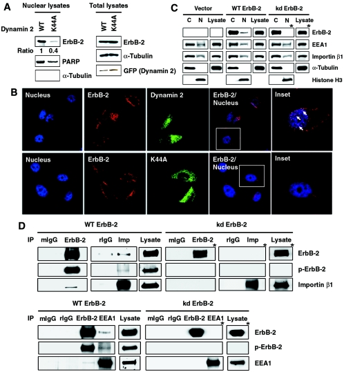
FIG. 8.
ErbB-2 nuclear translocation requires endocytosis. (A) Dynamin activity is required for nuclear localization of ErbB-2. CHO cells were transfected with ErbB-2 cDNA together with GFP-tagged WT or GFP-tagged K44A mutant (K44A) dynamin 2 cDNA. Left panels: the nuclear lysates from these cells were analyzed by Western blotting using antibodies against ErbB-2, PARP, and α-tubulin. The levels of ErbB-2 expression were quantitated and normalized to the protein levels of PARP. The expression level of ErbB-2 in GFP-tagged WT dynamin 2-transfected cells was set as 1. Right panels: total cell lysates were blotted for equal expression of ErbB-2, α-tubulin, and dynamin 2 (GFP). (B) CHO cells were transfected with the GFP-tagged WT (top) or K44A mutant (bottom) dynamin 2 together with ErbB-2 plasmid. Localization of ErbB-2 (red) in the TOPRO 3-stained nucleus (blue) was examined under a confocal microscope. ErbB-2 localized in the nucleus is shown in pink spots as indicated by arrows in the inset. (C) Cytoplasmic fractions (C), nuclear fractions (N), and total cell lysates from MCF-7/WT ErbB-2, MCF-7/kinase-deficient mutant ErbB-2 (kd ErbB-2), or vector control cells were analyzed by Western blotting with antibodies as indicated. (D) Lysates from MCF-7/WT ErbB-2 and MCF-7/kd ErbB-2 cells were immunoprecipitated (IP) with ErbB-2, importin β1 (Imp), EEA1, control mouse IgG (mIgG), and rIgG. The immunocomplexes were then analyzed by Western blotting with antibodies as indicated. Total cell lysates were also analyzed by Western blotting with anti-ErbB-2, antiphosphotyrosine (p-ErbB-2), anti-importin β1, and anti-EEA1 antibodies as positive controls. The blots marked with asterisks were exposed for a longer period.

