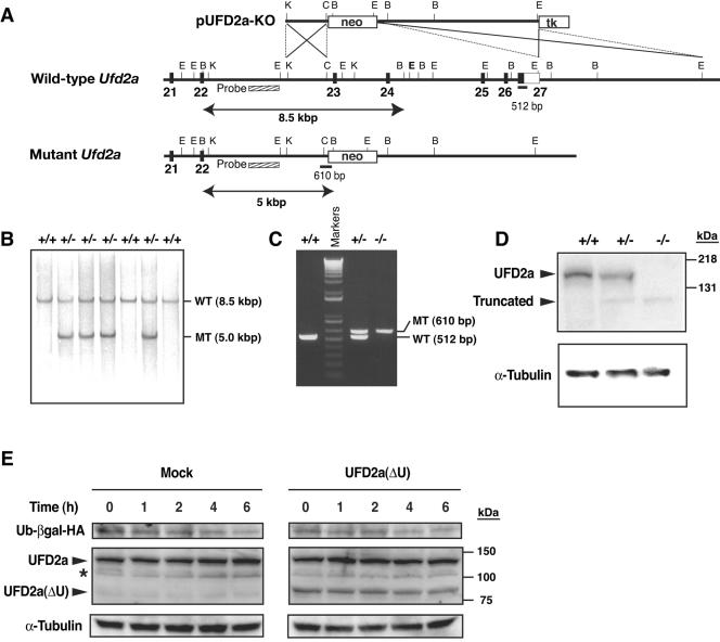
FIG. 1.
Targeted disruption of mouse Ufd2a. (A) Structures of the targeting vector (pUFD2a-KO), the mouse Ufd2a locus, and the mutant allele resulting from homologous recombination. The coding exons and coding portion of exon 27 are depicted by filled boxes, with the open box indicating the noncoding portion of exon 27. A genomic fragment used as a probe for Southern blot analysis is shown as a striped box, and the expected sizes of the BamHI fragments (arrows) that hybridize with the probe are indicated. The positions (solid bars) and sizes of PCR products used for screening are also indicated. neo, neomycin transferase gene linked to the PGK gene promoter; tk, thymidine kinase gene derived from herpes simplex virus linked to the PGK gene promoter. The orientations of both neo and tk are the same as that of Ufd2a. Restriction sites: E1, EcoRI; B, BamHI; C, ClaI; K, KpnI. Not all restriction sites are shown. (B) Southern blot analysis of genomic DNA extracted from the tail of adult mice. The DNA was digested with BamHI and subjected to hybridization with the probe shown in panel A. The positions and sizes of bands corresponding to the wild-type (WT) and mutant (MT) alleles are indicated, as are the genotypes of the analyzed mice. (C) PCR analysis of genomic DNA extracted from the yolk sacs of embryos of the indicated genotypes at E11.5. Amplification products corresponding to the black bars in panel A are indicated. (D) Immunoblot analysis of E11.5 embryo lysates with antibodies to UFD2a (top) and to α-tubulin (loading control) (bottom). The positions of full-length and truncated forms of UFD2a are indicated. (E) Cycloheximide chase analysis of the UFD substrate. HEK293T cells were transfected with an expression plasmid encoding Ub-βgal tagged with HA at its COOH terminus (Ub-βgal-HA) either alone (Mock) or together with a vector for UFD2a(ΔU). The cells were then treated with cycloheximide for the indicated times, lysed, and subjected to immunoblot analysis with anti-HA, anti-UFD2a, and anti-α-tubulin. The asterisk indicates a nonspecific band.

