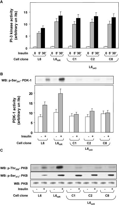
FIG. 3.
hIRΔ43 signaling in L6 skeletal-muscle cells. (A and B) L6, L6hIR, and L6Δ43 myoblasts (C1, C2, and C8 clones) were stimulated with 100 nM insulin for 10 min (B) or the indicated times (A) and then assayed for PI 3-K or PDK-1 activities as described in Materials and Methods. The bars represent the mean values plus standard deviations of data from three (A) and four (B) independent experiments, in triplicate. (B, top) Alternatively, the cells were lysed and Western blotted (WB) with specific phospho-Ser241 (p-Ser241) antibodies. The blots were revealed by ECL and autoradiography. The blot shown is representative of three independent experiments. (C) The cells were exposed to 100 nM insulin for 10 min and then solubilized as described in Materials and Methods. Cell lysates (50 μg of protein/sample) were blotted with specific phospho-threonine308-PKB antibodies (p-Thr308-PKB) and then reblotted with phospho-Ser473-PKB (p-Ser473-PKB) antibodies. To ensure equal Akt transfer, the filters were further blotted with PKB antibodies (PKB). The filters were revealed by ECL and autoradiography. The autoradiographs shown are representative of four independent experiments.

