
FIG.9.
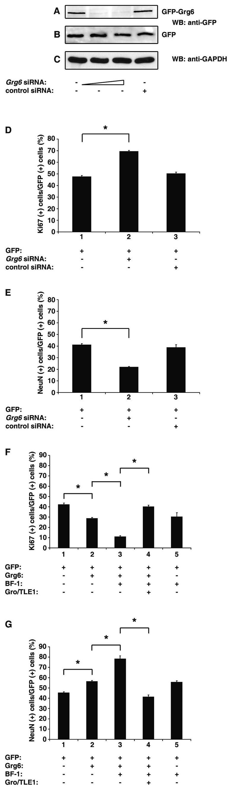
Involvement of Grg6 in cortical neurogenesis. (A to C) HEK293 cells were transfected with either GFP-Grg6 (A) or GFP alone (B) in the absence (lane 1) or presence of Grg6 siRNA (lane 2, 15 nM/transfection; lane 3, 30 nM/transfection) or control siRNA (lane 4, 30 nM/transfection). Forty-eight hours later, cell lysates were subjected to Western blotting (WB) analysis with anti-GFP (A and B) or anti-GAPDH (C) antibodies. (D and E) Primary cultures of mouse E11.5 to E12.5 cortical progenitor cells were transfected with GFP either alone (bar 1) or together with Grg6 (bar 2) or control (bar 3) siRNA (30 nM/transfection). Seventy-two hours later, cells were subjected to double-labeling analysis of the expression of GFP, Ki67, or NeuN. Results were quantitated as the percentage of GFP+ cells that were also positive for either Ki67 (D) or NeuN (E). The results are shown as the mean ± the standard deviation (*, P < 0.0001). (F and G) Primary cultures of mouse E11.5 to E12.5 cortical progenitor cells were transfected with either GFP alone (bar 1) or a combination of GFP and the indicated proteins (bars 2 to 5). Forty-eight hours later, cells were subjected to double-labeling analysis of the expression of GFP, Ki67, or NeuN and quantitation as described above (*, P < 0.0001).