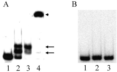
FIG. 2.
Gel shift assay. (A) The dofA promoter. The probe containing the region from nt −150 to −32 was mixed with FruA-DBD-His8, and the binding patterns were analyzed by PAGE. Lane 1, no FruA-DBD-His8; lane 2, 0.5 ng/μl of FruA-DBD-His8; lane 3, 1 ng/μl of FruA-DBD-His8; lane 4, 1 ng/μl of FruA-DBD-His8 and anti-FruA antibody. FruA-DBD-His8/dofA promoter complexes are indicated by arrows. Anti-FruA antibody/FruA-DBD-His8/dofA promoter complexes are indicated by an arrowhead. (B) The tps promoter. The probe contains the region from nt −250 to −41. Lane 1, no FruA-DBD-His8; lane 2, 0.5 ng/μl of FruA-DBD-His8; lane 3, 1 ng/μl of FruA-DBD-His8.

