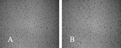
FIG. 4.
Images of polar and laterally attached bacteria. Cells of TR1-5 (csrA) were grown and allowed to attach to a coverslip for 4 h. A slide was assembled containing the cells at the upper surface (see Materials and Methods). Images (×640 magnification) of these cells were taken at the surface of the coverslip (A) or at 1.5 μm below the surface (B). The frame width for each image is 0.2 mm.

