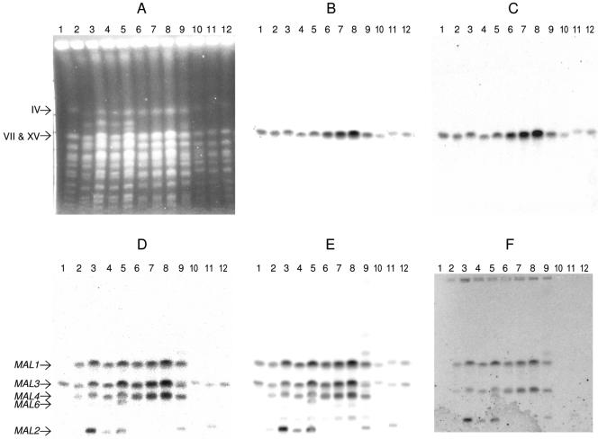
FIG. 3.
Detection of MAL loci. A PFGE gel was loaded with about 30 × 106 cells/lane. Strains were as follows: lane 1, Marker YNN 295; lane 2, A15; lane 3, A24; lane 4, A64; lane 5 A72; lane 6, A179; lane 7, A180; lane 8, A181; lane 9, A60; lane 10, S150-2B; lane 11, CEN.PK2-1D; and lane 12, RH144-3A. (A) Separated chromosomes stained with ethidium bromide. Chromosome IV and the duplex VII/XV are indicated. The gel was then blotted, and the blot was hybridized with the following probes: AGT1 (B), MAL13(AGT1) (C), MAL61 (D), MAL62 (E), and MAL63 (F). The MAL loci are identified on the left of panel D. The image for panel F was darkened to make the bands more visible.

