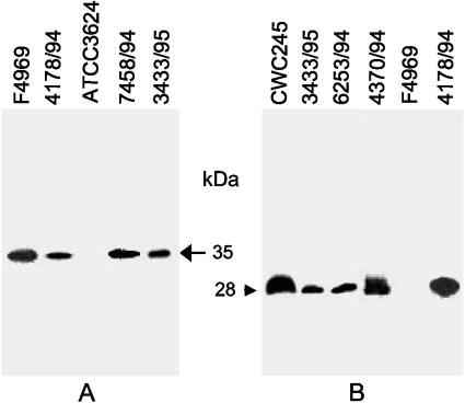
FIG. 5.
Western blot analysis of CPE and CPB2 production by selected SD isolates. (A) Western blot analysis of CPE production by selected SD isolates. C. perfringens strains were grown in DS medium and sonicated as described in Materials and Methods. An aliquot (25 μl) of each sonicated culture lysate was then subjected to SDS-polyacrylamide gel electrophoresis followed by Western blotting with CPE antibodies. The blot was developed by chemiluminescence detection to identify immunoreactive species. Results are shown for control strains F4969 (a plasmid cpe, non-food-borne GI disease isolate) and ATCC 3624 (a cpe-negative isolate) and for representative SD isolates 4178/94, 7458/94, and 3433/94. The arrow on the right indicates the migration of a CPE-specific immunoreactive band. (B) Culture supernatant proteins, prepared from each of the specified C. perfringens isolates, were subjected to SDS-polyacrylamide gel electrophoresis followed by Western blotting with CPB2 antibodies. The blot was developed by chemiluminescence detection to identify immunoreactive species. Results are shown for control strains CWC245 (a cpb2-positive, type C isolate carrying cpb2 on a large plasmid) and F4969 (a cpb2-negative, non-food-borne GI disease isolate) and for representative SD isolates 4178/94, 4370/94, 6253/94, and 3433/94. The arrow at the left indicates the migration of a CPB2-specific immunoreactive band.

