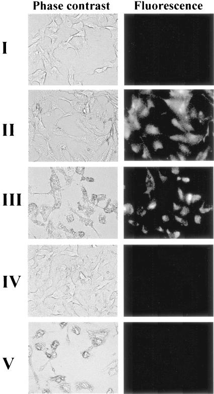
FIG. 1.
Visualization of infected or uninfected BGMK cells by use of MBs. Cells with or without permeabilization were incubated with 76μM of either MB CVB1/oligonucleotide hybrids (I and II) or MB CVB1 (III to V) for 1 h before being examined by phase-contrast and corresponding fluorescence microscopy. I, uninfected BGMK cells without permeabilization and with MB/oligonucleotide hybrids; II, uninfected BGMK cells with permeabilization and with MB/oligonucleotide hybrids; III, highly infected BGMK cells with permeabilization and with MBs; IV, uninfected BGMK cells with permeabilization and with MBs; V, highly infected BGMK cells with permeabilization and with nonspecific MBs.

