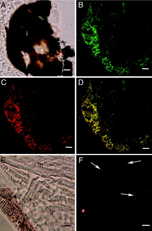
FIG. 5.
Fluorescence in situ hybridization of ant lion cryosections. A. Transmitted-light micrograph of a cryosection through the abdomen of an ant lion showing the dark pellet of indigestible material at the barrier between the midgut and the hindgut (i). B to D. Corresponding confocal micrographs of FISH using EUB338 (green, panel B) and probe D (red, panel C) and overlay showing EUB338 and probe D (panel D). Yellow objects associated with the undigested material are hybridizing with EUB338 and probe D (panel D). E. Transmitted-light micrograph of a cryosection through the abdomen of the ant lion showing adipose (a) and muscle (m) tissue near the body wall. D. Corresponding confocal micrograph of FISH using EUB338 (green) and W2 (red). Yellow-orange objects scattered in tissue (arrows indicate examples) are hybridizing with EUB338 and W2. For A to D, scale bars represent 20 μm; for E and F, scale bars represent 10 μm.

