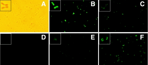
FIG. 2.
Fluorescent A. nicotinovorans cells upon gfp expression. (A) Light microscopy image of A. nicotinovorans bacteria. Inset, magnified individual cell field. (B) Fluorescence microscopy image of A. nicotinovorans transformed with pART2-gfp; (C) A. nicotinovorans/pAO1 transformed with pART2-gfp; (D to F) A. nicotinovorans/pAO1 transformed with pART3-gfp and grown in the absence of nicotine (D), in the presence of nicotine for 60 min (E), and in the presence of nicotine overnight (F). Bacteria shown in panels A to C were grown in LB medium, and bacteria shown in panels D to F were grown in citrate medium supplemented with 0.5% yeast extract, mineral salts (5), and, as required, 0.05% l-nicotine. Pictures of A. nicotinovorans bacteria were taken using an Zeiss Axioskop 50 epifluorescence microscope equipped with a Plan-Nofluar 100× (1.3-numerical-aperture) objective and a triple-pass filter set. Digital images were recorded with a Nikon D100 camera.

