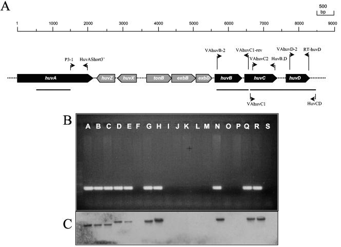
FIG. 2.
(A) Physical map of the heme uptake cluster of V. anguillarum O1 strain 775 (see reference 20). Thick arrows denote ORFs, and the scale above indicates the length in nucleotides. Thin arrows indicate the position and direction of primers used in the PCR screening and in the amplification of huvB and huvCD DNA probes. Horizontal bars denote location of DNA probes. (B) Results of PCR screening for presence of the heme receptor huvA gene in a collection of V. anguillarum strains. (C) Southern blot confirmation of presence of huvA gene. Letters in panels B and C denote V. anguillarum strains as follows: A, 775; B, R-82; C, TM-14; D, 96-F; E, ATCC 14181; F, ATCC 43306; G, RV22; H, 43-F; I, PT-493; J, 13A5; K, 11008; L, ET-208; M, ATCC 43307; N, RPM 41.11; O, ATCC 43310; P, ATCC 43311; Q, ATCC 43312; R, ATCC 43313; S, ATCC 43314.

