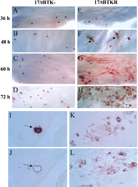
FIG. 4.
Lytic viral protein expression in 17/tBTK−-infected (A to D) and 17/TBTKR-infected (E to H) ganglia 36 h (A and E), 48 h (B and F), 60 h (C and G), and 72 h (D and H) p.i. using WGIHC as described in Materials and Methods. In 17/tBTK−-infected ganglia, lytic viral protein-expressing neurons remain discrete with no evidence of lateral spread. In contrast, spread of virus in TGs of 17/tBTKR-infected mice is apparent by 48 h p.i. (arrows). Panels I to L show serial sectioned frozen ganglia from 17/tBTK− (I and J)- and 17/tBTKR (K and L)-infected mice harvested on day 4 day p.i. and immunostained for ICP27 (I and K) and gC (J and L). Arrows in panels I and J indicate neurons containing detectable ICP27 but no gC (dashed line indicates cell boundary).

