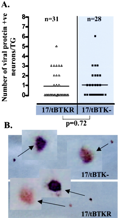
FIG. 8.
(A) Comparison of the number of neurons expressing IE proteins in TGs infected with 17/tBTK− (squares) and 17/tBTKR (triangles) 30 h p.i. TGs were probed with antibodies directed against ICP27 and ICP0 as described in Materials and Methods. The difference between the groups was not significant (Student's t test). (B) Positive neurons representative of the intensity of staining in 17/tBTK−- and 17/tBTKR-infected ganglia.

