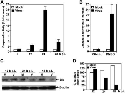
FIG. 4.
Activation of caspase-8 and cleavage of proapoptotic protein Bid by MHV infection. (A) Caspase-8 activity. Cells were infected with MHV at an MOI of 5. At various time points p.i. as indicated, cell extract was prepared, and cleavage of pro-caspase-8 and the presence of the active caspase-8 were detected with a caspase colorimetric protease assay kit using specific substrates as described in Materials and Methods. The caspase-8 activity from virus-infected cells was expressed as means ± standard error from three independent experiments and as fold increase over that from mock-infected control, which was set as onefold. (B) Effect of caspase-8 inhibitor II on caspase-8 activity. Cells were treated with cell-permeable caspase-8 inhibitor II (C8-inh.) 1 h prior to and during virus infection for 24 h or with DMSO as a negative control. Mock-infected cells were used for normalizing the caspase-8 activity expressed as fold increase as described for panel A. (C) Bid cleavage. Cells were infected with MHV or mock infected with PBS. Cell lysates were collected at various time points p.i. as indicated. The protein level for the full-length Bid was detected by Western blotting using a Bid-specific antibody. The housekeeping protein β-actin was used as an internal control. (D) Quantification of protein bands shown in panel C. The protein bands were quantified by densitometric analysis with UVP software, normalized to β-actin, and expressed as percent relative abundance to Bid of mock-infected cells at 12 h p.i., which was set as 100%.

