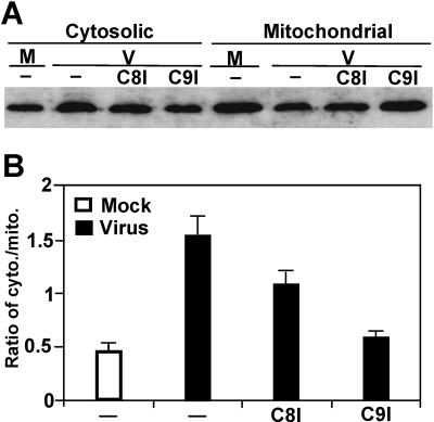
FIG. 5.
Effect of caspase-8 and -9 inhibitors on cytochrome c release during MHV infection. (A) Cells were treated with caspase-8 or -9 inhibitors 1 h prior to and throughout the infection. At 48 h p.i., cells were lysed. The cytosolic and mitochondrial fractions were separated by differential centrifugation. Cytochrome c was detected by Western blotting with a specific antibody as described in the legend to Fig. 2. M, mock infection; V, virus infection; C8I and C9I, caspase-8- and -9 inhibitor, respectively; −, no treatment with caspase inhibitor. (B) Quantification of the protein bands shown in panel A and two additional gels (not shown). The amount of cytochrome c was quantified by densitometric analysis with UVP software. The efficiency of release of cytochrome c from mitochondria into cytosol is expressed as a ratio of cytosolic fraction to mitochondrial fraction and as means ± SD.

