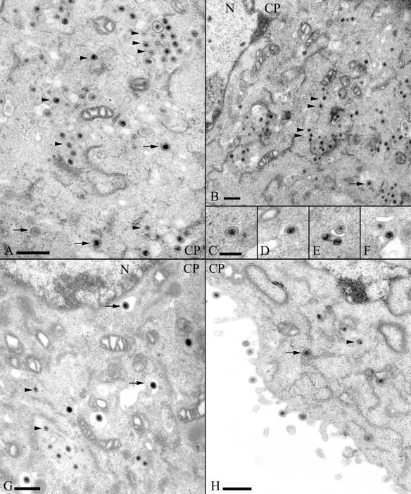
FIG. 4.
Electron micrographs of Rk13 cells infected with L11Δ20 (A to F) or with L11Δ20R (G to H) (both at 16 h p.i.). Different kinds of particles are demonstrated within the cytoplasm of cells (CP), and some enveloped particles are designated by arrows, whereas arrowheads point to examples of unenveloped cytoplasmic capsids. Inlays C to F depict particles during “budding,” as frequently observed in cells infected with L11Δ20. Bars represent 500 nm in A, B, G, and H and 250 nm in C.

