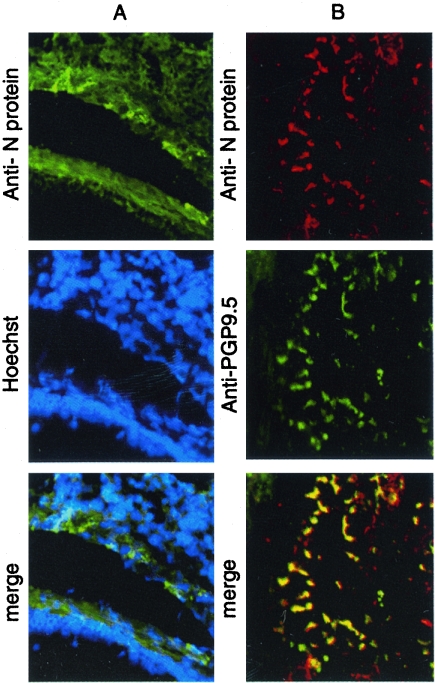
FIG. 4.
Codetection of RSV N protein and neuronal marker PGP9.5 in RSV-infected lung tissue. (A) RSV N protein was detected primarily in the epithelial walls surrounding small bronchioles (longitudinal section), and Hoechst staining shows only a few RSV-positive cells (most likely epithelial cells). (B) RSV N protein staining overlapped with neuronal marker PGP9.5 in nervous fibers in a small bronchiole (transversal section). These nerve fibers may come from one neuron (Magnification, ×32).

