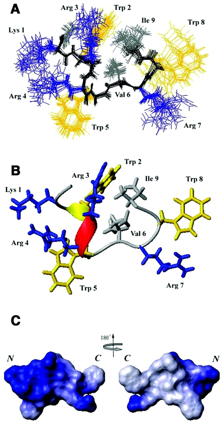
FIG. 5.
Calculated structures of Pac-525. (A) Twenty lowest-energy structures calculated for Pac-525 (2 mM in 20 mM sodium phosphate buffer at pH 4.5 and 37°C with 200 mM SDS). (B) The final refined average structure for Pac-525 bound to SDS. The positively charged residues are shown in blue, the tryptophan residues are shown in yellow, and the hydrophobic residues are shown in gray. (C) The electrostatic surface plot of Pac-525. Positive charge is indicated by blue and neutral charge is indicated by white. The figure was generated using the program MOLMOL (21).

