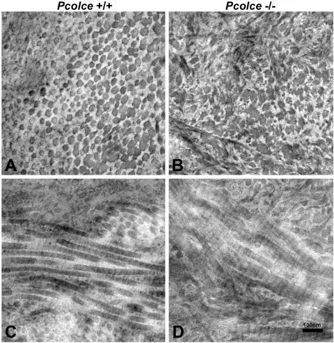
FIG. 4.
Transmission electron micrographs of collagen fibril morphology in 4-week-postnatal Pcolce−/− and wild-type (Pcolce+/+) left femora. Unlike wild-type fibril cross sections (A), all mutant collagen fibrils have irregular scalloped profiles in cross section (B), whereas longitudinal views of collagen fibrils (C and D) show Pcolce−/− fibrils to have irregular, broadened profiles, with some branching.

