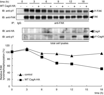
FIG. 7.
Kinetic changes in the level of FAK tyrosine phosphorylation by CagA. AGS cells transfected with WT CagA-HA expression vector or control empty vector were harvested at the indicated time points after transfection. Cell lysates were immunoprecipitated with anti-FAK antibody or normal rabbit IgG. Immunoprecipitates (IP) and total cell lysates were immunoblotted (IB) with the indicated antibodies. Arrows indicate positions of FAK, tyrosine-phosphorylated FAK (pY-FAK), and tyrosine-phosphorylated CagA (pY-CagA) (top). Quantitation expressed as the ratio of tyrosine-phosphorylated FAK to total FAK is summarized in the graph on the bottom. Each value was calculated from the intensities of anti-pY and anti-FAK immunoblotting by using a luminescence image analyzer and defining the value in the untransfected AGS cells (time 0) as 100%.

