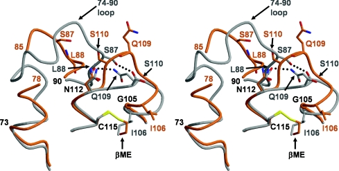
Figure 4. The loss of two hydrogen bonds as a result of the shift in the SXN motif contributes to disordering of the 74–90 region in Cys115-modified PBP 5.
In this stereo view, wild-type PBP 5 is represented by a grey backbone and side chains, whereas 2ME-modified PBP 5 is shown in orange. In wild-type PBP 5, there are two potential hydrogen bonds (shown as dashed lines) that link the SXN motif with the 74–90 region, which is fully ordered. In 2ME-modified PBP 5, these bonds are absent due to the shift in residues 106–111 induced by the 2ME alkylating Cys115, and their loss is hypothesized to lead to destabilization and disordering of the 74–90 loop. Residues that differ significantly in position between the two structures are coloured grey for wild-type and orange for 2ME-modified PBP 5. This Figure was prepared with MOLSCRIPT [47] and RASTER3D [48].

