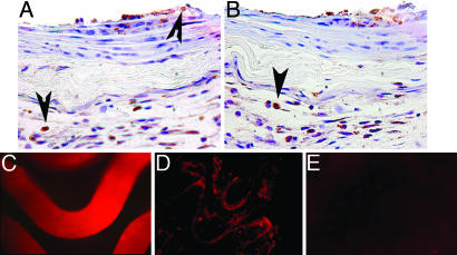
Fig. 4.
PAA-BP-modified steel stents: Inflammatory response and tissue distribution of the vector in vivo. Representative DAB-immunohistochemistry photomicrographs, demonstrating prevalence of CD68-positive macrophages (indicated by the arrowheads) in arterial sections treated with bare metal (A) and PAA-BP-modified (B) stents (original magnification, ×200). Fluorescent photomicrograph of a Cy3-Ad-modified stent surface (2.5 × 10⁹ viral particles per stent) before deployment (C) and its imprint (en face) on the luminal surface of a rat carotid artery (D) 24 h after stenting. (E) Absence of autofluorescence in a rat carotid artery stented without tethered Ad. (C–E) Original magnification is ×200, rhodamine filter set.

