Fig. 2.
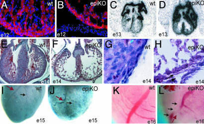
Histologic and morphologic analysis of epicardial mutant RXRα embryos. (A and B) A thin myocardium is detected as early as E12 and corresponds to decreased number of myocytes, as indicated by staining with the muscle marker sarcomeric α-actinin (red, α-actinin; blue, DAPI nuclear staining). (C and D) Epicardial RXRα mutation is sufficient to impair the normal down-regulation of mlc2a in E13 ventricles, and reflects deficient specification in the mutant myocardium. (E and F) Epi-RXRα mutant embryos at E14 display a lack of myocardial compaction, as indicated by hematoxylin–eosin staining. (G and H) E14 epi-RXRα mutant ventricles also exhibit epicardial detachment and thickened subepicardial space, suggesting deficient formation and/or activation of the epicardial epithelium (arrows). (I–L) Epi-RXRα mutant ventricles at E15 present altered coronary arteriogenesis, with tortuous vessels (J, red arrow) and defective branching (J, black arrow) as shown by whole-mount immunohistochemistry using a Pecam-1 antibody. (K and L) Photographs of freshly dissected E16 hearts.

