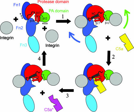
Fig. 4.
Cartoon showing hypothesized effect of integrins (gray) on SCPB structure. Domains of SCPW are indicated and colored as in Fig. 1. Positions of RGD sequences in PA domain and Fn1 are indicated as are the β12, β17, and β18 strands. In step 1, binding of integrins to RGD sequences disrupts the β12–β17 interactions, allowing the β17–β18 hairpin to rotate and promote formation of helix hC. Fn2 is pulled back, making space for C5a binding in step 2. In step 3, C5a is proteolyzed. In step 4, the integrins dissociate regenerating free SCPB.

