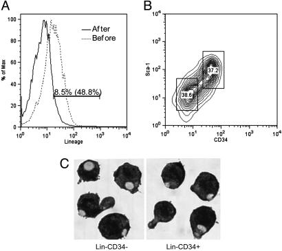
Fig. 1.
Isolation of Lin–CD34+ and Lin–CD34– EML cells. (A) Expression of the lineage markers on EML cells before lineage depletion (dotted line) or after lineage depletion (solid line) by magnetic cell sorting. The cells were stained with phycoerythrin (PE)-cy7-lineage mixture antibodies and analyzed by FACS. (B) Lin–-enriched EML cells were stained with PE-cy5-lineage mixture antibodies, PE-conjugated CD34 antibody, and allophycocyanin-conjugated Sca-1 antibody. Subsequently, the Lin+ cells were gated out and Lin– EML cells were sorted into CD34+ and CD34– EML cell populations by Moflo high flow cell cytometry. (C) Lin–CD34– and Lin–CD34+ EML cells cytospun onto slides were stained with Wright-Giemsa by standard protocols. (Magnification: ×400.)

