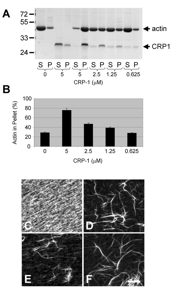
Figure 4.
CRP1 bundles F-actin independently. The indicated concentration of CRP1 was incubated with actin filaments for 30 min at room temperature and centrifuged at 10,000 × g. Proteins from the supernatant (S) and the pellet (P) were separated by electrophoresis and detected by Gelcode Blue staining (A). The percentage of total actin in the pellet was quantified as described in Experimental Procedures (B). n = 4 ± SEM. Bundling reactions were also carried out on glass coverslips, fixed, and stained with rhodamine-phalloidin to allow visualization of the actin filaments bundles: (C) actin filaments, (D) actin filaments + 2.5 μM α-actinin, (E) actin filaments + 5.0 μM CRP1, and (F) actin filaments + 2.5 μM α-actinin + 2.5 μM CRP1. Results are representative of 4 separate experiments. Bar = 10 μm.

