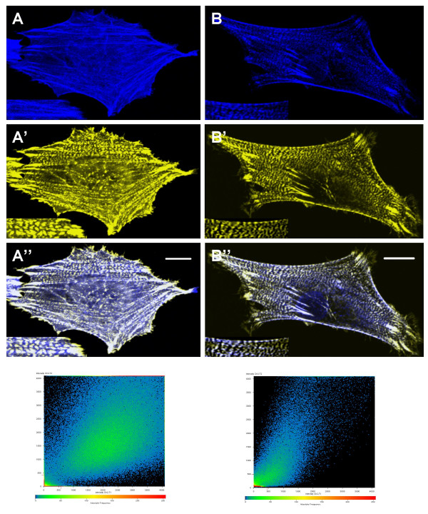
Figure 5.
Two populations of CRP1 are localized along stress fibers. REFs expressing CFP-CRP1 and YFP-α-actinin were fixed with 3% formaldehyde in PBS (A) or Triton X-100 buffer (B) and prepared for confocal microscopy as described in Experimental Procedures. Images of CFP-CRP1 (A and B) and YFP-α-actinin (A' and B') localization are shown. A zoomed region is shown in the lower left corner of each image. Merged images are shown in A" and B". Results are representative of 4 separate experiments. Bar = 10 μm.

