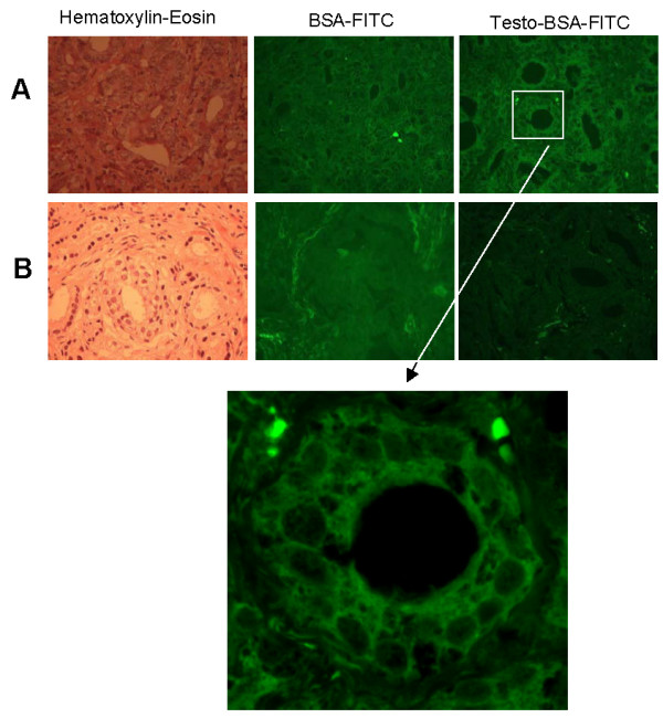
Figure 2.

Representative cases of mAR positive (A) and negative (B) tumor specimens. A representative case of mAR positive (A) and a negative case (B) are shown. Left panels present H&E staining, middle panels present BSA-FITC staining, while right panels present staining with testosterone-BSA-FITC. Note the membrane localization of fluorescence in positive cells. Lower panel presents a higher magnification of the square region shown. In all cases preincubation with 3% BSA and 10-4M cyproterone acetate was performed, prior to mAR detection. Compare Figure 2 with Figure 1 (no preincubation).
