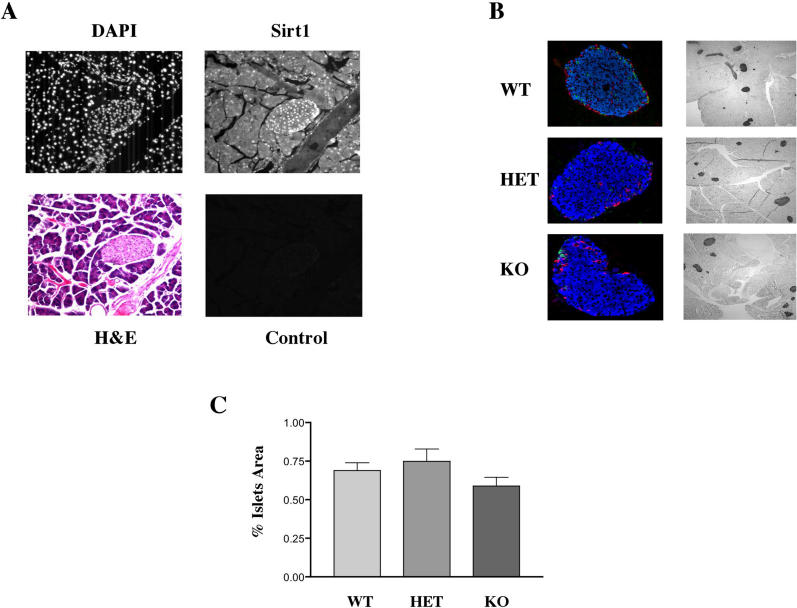
Figure 1. Sirt1 Is Localized in the Islets of Langerhans.
The pancreas of wild-type mice was sectioned and stained as described.
(A) Nuclear staining using DAPI (top left). Immunofluorescence using Sirt1 antibody (top right); hematoxylin and eosin staining of the same section of pancreas (bottom left); immunofluorescence control using a rabbit secondary antibody (bottom right).
(B) Pancreases of wild-type (WT), Sirt1+/− heterozygotes (HET), or Sirt1−/− homozygous KO mice were stained with antibodies against insulin (blue), glucagon (red), or somatostatin (green) (shown in left column). Representative islets of mice of all three genotypes are shown. Pancreases were also silver-stained for morphometry (right column). Islets appear as dark figures and their area was determined by scanning, using Image-Pro 4.1 Plus software.
(C) The areas are shown as percentage of area of the entire pancreas.

