Figure 4.

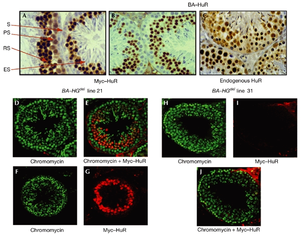
Analysis of endogenous and transgenic HuR expression at the cellular level. (A–C) Immunohistochemical analysis; testis sections (3–5 μm) from β-actin–HuR (BA–HuR) lines 19 (A) and 18 ((B) and (C)) were incubated with the 9E10 antibody to detect the HuR transgene ((A) and (B)), or with the 19F12 antibody to detect endogenous HuR (C). (D–J) Confocal analysis. Testis sections (∼60 μm) from BA–HGdel lines 21 (D–G) and 31 (H–J) were incubated with chromomycin (green), which specifically stains nuclei (D, F, H), or with 9E10 (red), which detects transgenic HuR (G, I). Double labelling gave an orange staining, which was mainly restricted to the round spermatid population; this was only visible in BA–HGdel line 21 sections (E), and not in BA–HGdel line 31 sections (J). ES, elongated spermatids; PS, primary spermatocytes; RS, round spermatids; S, spermatogonia.
