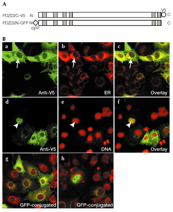
Figure 1.

Subcellular localization of PDZD2. (A) The PDZD2C–V5 and PDZD2N–GFP constructs, containing a carboxy-terminal V5 tag and an amino-terminal green fluorescent protein (GFP) tag, respectively. Shaded boxes indicate the positions of the PDZ domains. (B) Localization of PDZD2 in COS7 cells. Cells transfected with PDZD2C–V5 (a–f) were immunostained with an anti-V5 antibody and counterstained with concanavalin A (conA) to stain the endoplasmic reticulum (ER) (b) or propidium iodide to detect nuclear DNA (e). Superimposed images are shown in c and f. Arrows indicate the co-localization of V5-tagged PDZD2 with the ER marker, and arrowheads indicate nuclear localization of PDZD2 (seen in ∼8% of the transfected cells). Fluorescent images of PDZD2N–GFP-transfected cells that display ER and nuclear staining, respectively, are shown in g and h; these cells were counterstained with conA.
